UPMC 3D Printing Program
Program overview
Traditional medical images are flat: surgeons mentally reconstruct three-dimensional anatomy from a stack of two-dimensional slices. A 3D-printed model removes that guesswork. Holding a true-to-scale replica of a patient's anatomy gives the care team an instant, intuitive understanding of complex problems, lets them plan and even practice key steps of a procedure beforehand, and (for sterilizable models) provides a reference that can be brought right into the operating room.
The program began in 2016 with a single resident and two 3D printers, funded by a Chairman's grant from Interventional Radiology and a Beckwith Institute Frontline Innovation Grant. Its founding goal was simple: create low-cost, high-quality 3D-printed models to improve procedure planning and reduce costs. Today it is a fully resourced clinical service, with a 4.4 full-time-equivalent team of biomedical engineers serving physicians across the health system from a dedicated facility built around ISO 7 clean rooms.
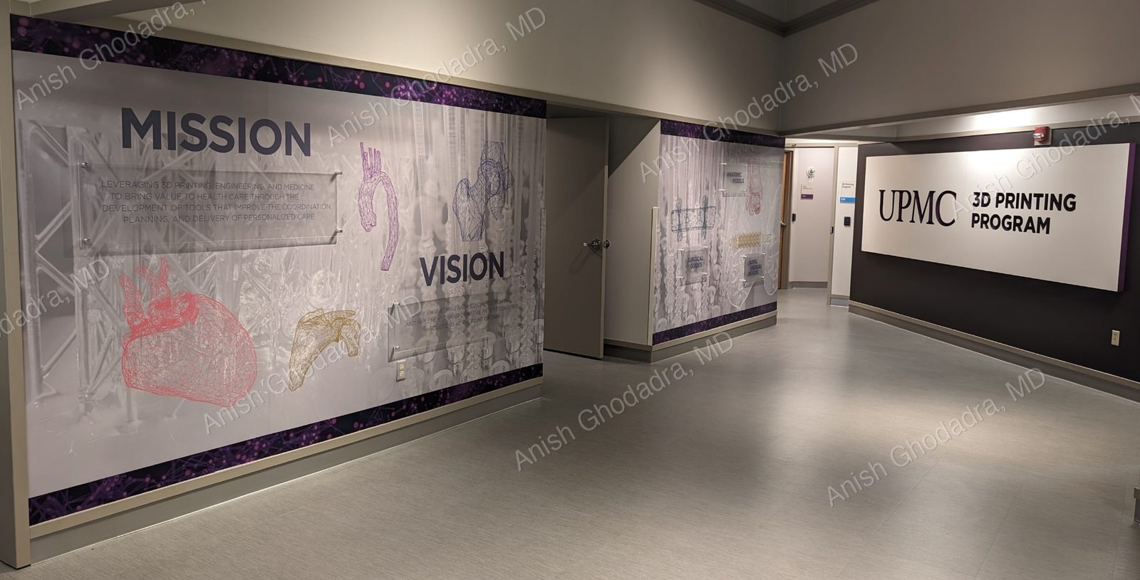
Vision
Unify engineering and medicine to design and implement transformative patient-centered solutions at the point of care.
Mission
Leveraging 3D printing, engineering, and medicine to bring value to health care through the development of tools that improve the coordination, planning, and delivery of personalized care.
Clinical applications
Anatomic models account for roughly 90% of the program's work, but the same engineering capability supports a growing range of clinical uses.
Patient-specific anatomic models
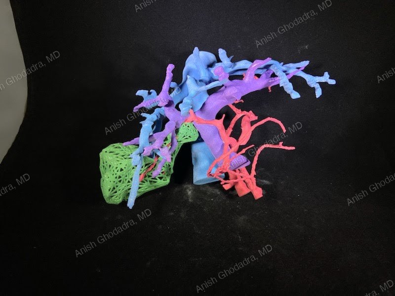
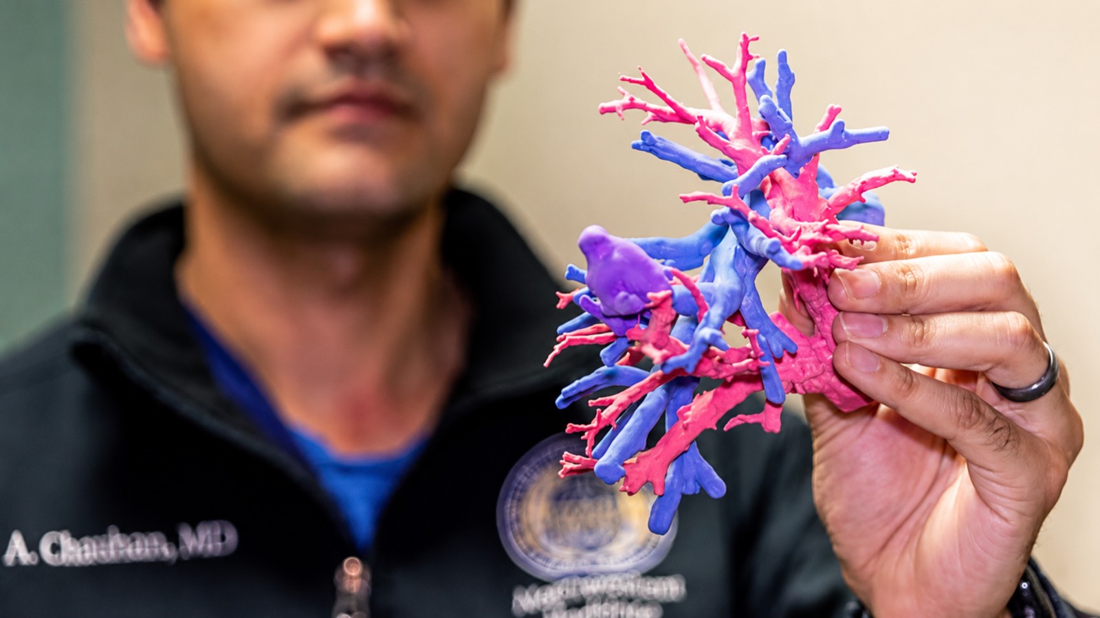
Built from a patient's own imaging, these models let surgeons visualize complex or rare anatomy before they ever make an incision. Published analyses have shown that anatomic models and guides can reduce operating room time in orthopaedic and maxillofacial surgery, translating directly into cost savings and improved efficiency.
Surgical rehearsal and simulation
For difficult approaches, such as endoscopic endonasal surgery, physical models allow the team to rehearse the procedure on the exact anatomy they will encounter. Models are also used for resident training, research, and education.
Sterilizable intra-operative models
Certain materials can be sterilized and autoclaved, so a model can be brought into the sterile field and referenced during the operation itself, not just beforehand.
Virtual surgical planning and 3D-printed surgical guides
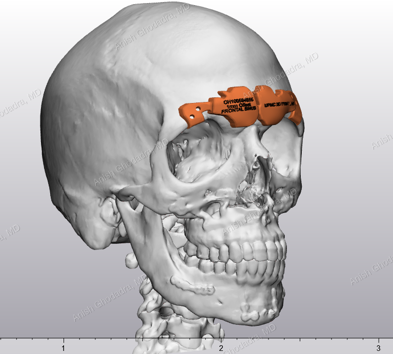
Beyond models, the program produces patient-specific surgical guides that translate a digital surgical plan into the operating room. In mandibular reconstruction, for example, virtual surgical planning maps the tumor resection, the fibular graft harvest, and the graft placement, and 3D-printed cutting guides reproduce that plan precisely on the patient. In orbital floor reconstruction, a mesh plate that would normally be bent by hand can instead be pre-bent against a printed model, reducing operative time and improving symmetry. Mirroring techniques, printing the healthy side as a template for the injured side, help restore normal anatomy in trauma reconstruction.
In the news: “jaw in a day”
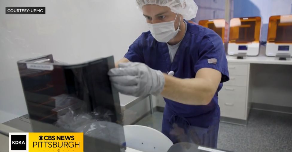
In September 2025, KDKA CBS News Pittsburgh followed a patient through the kind of reconstruction this planning makes possible. Andy Potocki, a science teacher, hockey player, and wedding DJ from Erie, had an ameloblastoma: a tumor growing through his jawbone, pushing his teeth out of place and wrapping around nerves. In a six-hour operation, Dr. Matt Spector removed the diseased section of jaw and rebuilt it with bone taken from Potocki's own fibula.
The lab built an exact replica of Potocki's jaw from scans of his face and leg, so the team could work out in advance where to cut and where the dental implant sockets would sit. Those sockets were drilled into the leg bone before it was ever taken out of the leg. “The 3D modeling and the engineering behind it is what's really been the big step forward,” Spector told KDKA.
The approach is known as “jaw in a day.” What once took several surgeries spread over six months to a year is now largely done in one: the patient wakes up with the jaw rebuilt and their dental structures intact, with the implants themselves fitted at later visits.
Read and watch the full story at CBS News Pittsburgh
3D printing technologies
The program operates several complementary printing technologies, chosen per case based on the anatomy, material properties, and intended use.
| Technology | How it works | Strengths |
|---|---|---|
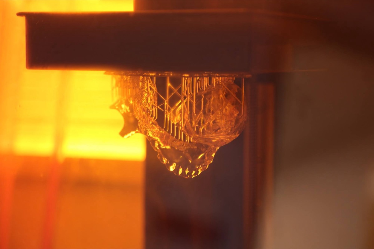
| Stereolithography (SLA) | A UV laser hardens liquid resin one thin layer at a time. | High precision and fast print times; clear, rigid, translucent-elastic, silicone, and biocompatible, autoclavable materials. |
| MultiJet Fusion (MJF) | Selectively melts and colors PA12 nylon powder. | Full-color models, complex geometries, and durable nylon parts. |
| Fused Deposition Modeling (FDM) | Extrudes melted thermoplastic filament layer by layer. | Multiple colors and materials with low-cost printers and materials. |
| Polymer Jetting (PolyJet) | Jets and cures fine droplets of photopolymer. | Clear and multi-color models with biomimetic, tissue-like materials. |
ISO 7 clean rooms
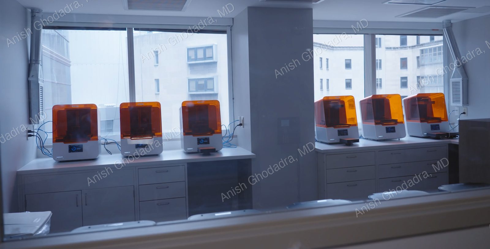
Printing does not happen on a bench in a back office. The program builds its models inside ISO 7 clean rooms, a classified environment in which airborne particles are held to a defined limit through filtered air, a high rate of air exchange, controlled surfaces and materials, and gowning before entry.
That environment matters for point-of-care manufacturing. Liquid resins and nylon powders pick up whatever is in the air around them, and airborne contamination during a build shows up later as surface defects, weak layers, or a part that cannot be cleaned properly. Controlling the room is what allows the program to produce models to a documented, repeatable standard, including the sterilizable models that are autoclaved and carried into the sterile field.
From image to 3D-printed model
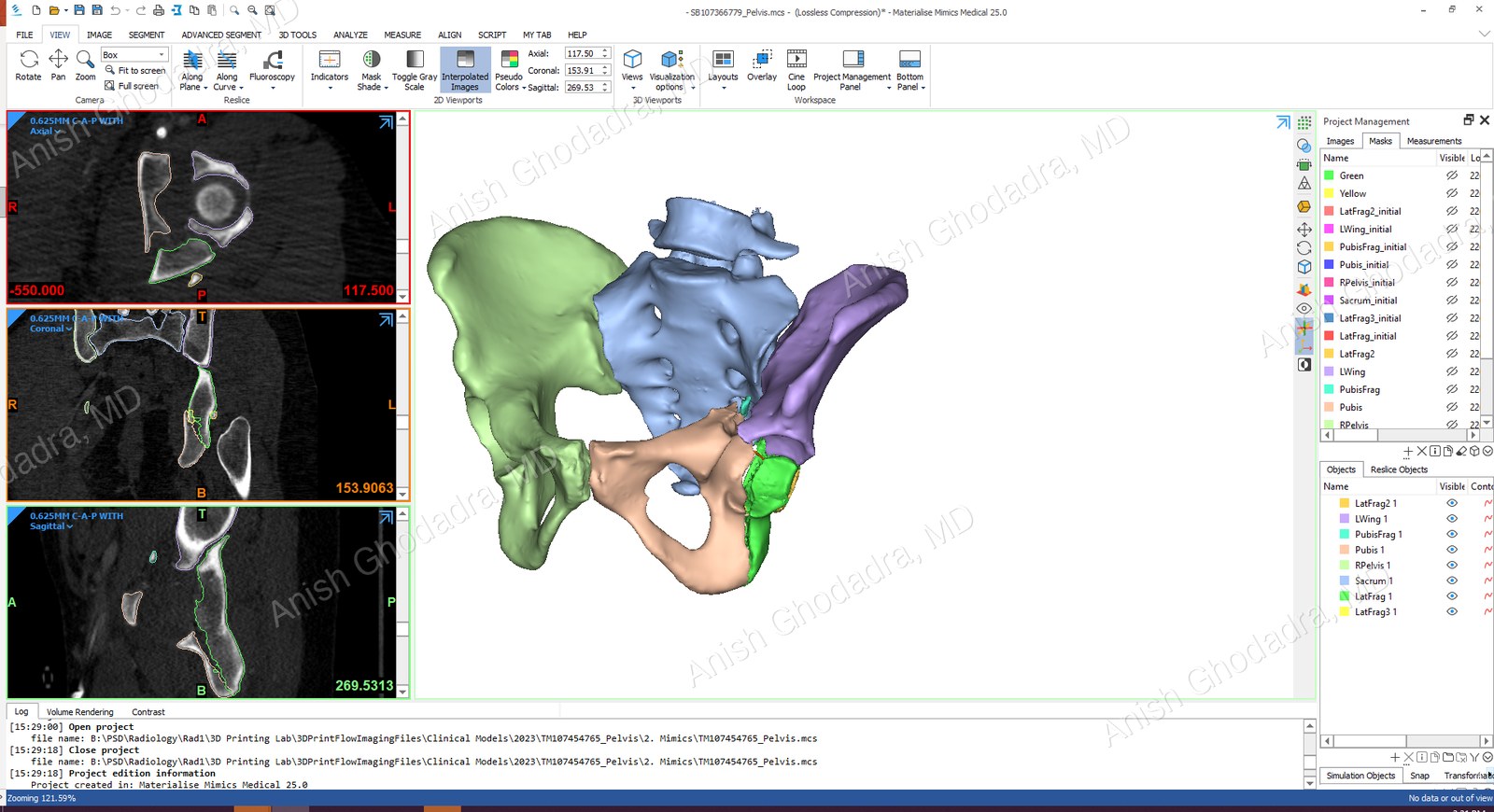
Every model follows a controlled, documented manufacturing pathway:
- Imaging: A diagnostic CT, MRI, or ultrasound is acquired or sourced.
- Segmentation: The relevant anatomy is traced out slice by slice, a careful step that takes anywhere from 10 minutes to 3 hours and must be as accurate as possible.
- STL generation: The segmented anatomy is converted into a printable 3D mesh.
- CAD and modification: The model is refined and modified, and any guides or fixtures are designed.
- Print preparation: The optimal printer, material, and build orientation are selected, and support structures are generated.
- Printing: The model is produced on the chosen technology.
- Post-processing: Supports are removed, parts are post-cured, cleaned (including bead-blasting excess nylon powder), assembled, and colored.
- Quality control and documentation: Each model is inspected and fully documented before release.
The entire workflow is tracked in PrintFlow, a custom UPMC-developed software solution that provides full traceability throughout the manufacturing process. The underlying technology was awarded a patent in 2024.
Custom devices & innovation
The same engineering capability is regularly turned to solving practical clinical problems, replacing broken, discontinued, or unavailable equipment, often at a fraction of the original cost.
| Solution | Problem solved | Impact |
|---|---|---|
| Gamma Knife head cradle | An adjustable MRI head cradle for Gamma Knife patients, broken beyond repair and no longer manufactured. | Improved design replaced a ~$1,000 device; easier positioning and better throughput. |
| Gamma Knife fiducial box | A posterior plate not sold in parts, with requested design changes. | Redesigned in-house, replacing another ~$1,000 device. |
| Apnea Link case | A detachable case for sleep-apnea study devices whose handles broke beyond repair and were out of warranty. | Used on 50+ devices, drastically improving study efficiency. |
| MEG glasses | Fragile glasses worn during MEG brain-mapping procedures, broken beyond repair and halting non-emergent mapping. | Improved design filled a supply-chain gap and recovered ~$300 per pair. |
| MRI head coil bracket | A platform that holds a patient's head in the MRI coil, with a persistent non-serviceable part failure. | Redesign enabled serviceability and avoided a $60,000–$90,000 coil replacement. |
| COVID-19 nasopharyngeal swab | A worldwide shortage of test swabs in early 2020. | Designed and in production within 3 weeks in a converted OR; UPMC registered as an FDA medical device manufacturer, and 62,000+ patients were tested across 20+ Pennsylvania sites. |
Milestones & recognition
- 2016: Program established with a Chairman's grant from Interventional Radiology and a Beckwith Institute Frontline Innovation Grant.
- 2019, Capitol Hill: At the 10th annual Medical Imaging Technology Showcase in Washington, DC, the program was highlighted as an example of the importance of radiology and imaging in patient care. A parent, Stacey Matvya, told lawmakers how a 3D-printed model of her young daughter Charlee's kidneys helped the family understand the disease and helped surgeons save the kidney.
- 2020, COVID-19 response: The team developed an FDA-registered 3D-printed nasopharyngeal swab within three weeks, ultimately supporting testing of more than 62,000 patients across the region.
- 2024, Patent: A patent was granted for the technology behind PrintFlow, the program's custom workflow and traceability software.
- 2025, In the news: KDKA CBS News Pittsburgh featured the lab's role in “jaw-in-a-day” mandibular reconstruction, following an Erie patient from tumor to rebuilt jaw.
- Today: A dedicated new facility, a 4.4 FTE engineering team, and national recognition as a leader in point-of-care 3D printing.
Leadership & team
-
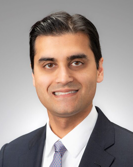 Anish Ghodadra, MD Medical Director
 -
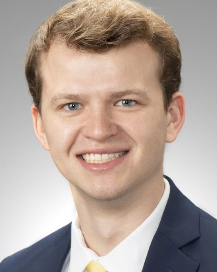 Sean Graves, BS Lead Biomedical Engineer
 -
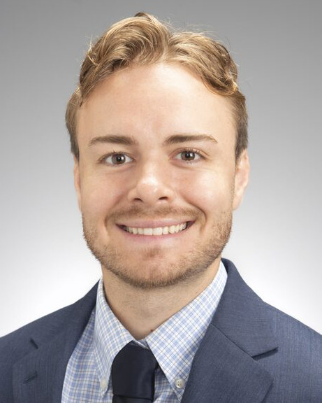 AJ Cook, MS Intermediate Biomedical Engineer
 -
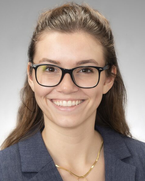 Jadan Law, BS Associate Biomedical Engineer
 -
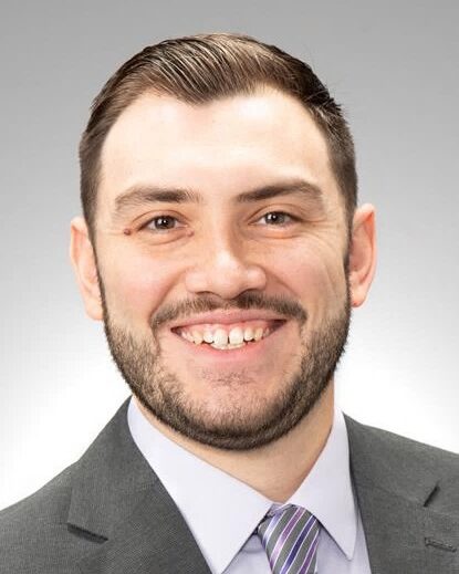 Nicholas Torres, BS Associate Biomedical Engineer
Anish Ghodadra, MD, the program's founder and Medical Director, is an interventional radiologist and Associate Professor of Interventional Radiology and Bioengineering at the University of Pittsburgh. He has co-authored multi-society RSNA Special Interest Group consensus guidelines on the appropriate clinical use of 3D printing in radiology. (Learn more about Dr. Ghodadra.)
Access & cost
- For patients: 3D-printed models are currently covered by UPMC, with no added cost to the patient. If 3D printing may help in your care, ask your UPMC physician whether a model or surgical guide is appropriate.
- For referring physicians: The program accepts requests from across UPMC's specialties. Learn more at upmc.com/3dprinting or contact the program directly at ghodadraa@upmc.edu.
- UPMC imaging line: 412-647-8762 or 800-533-8762.