The
Ghodadra
Lab
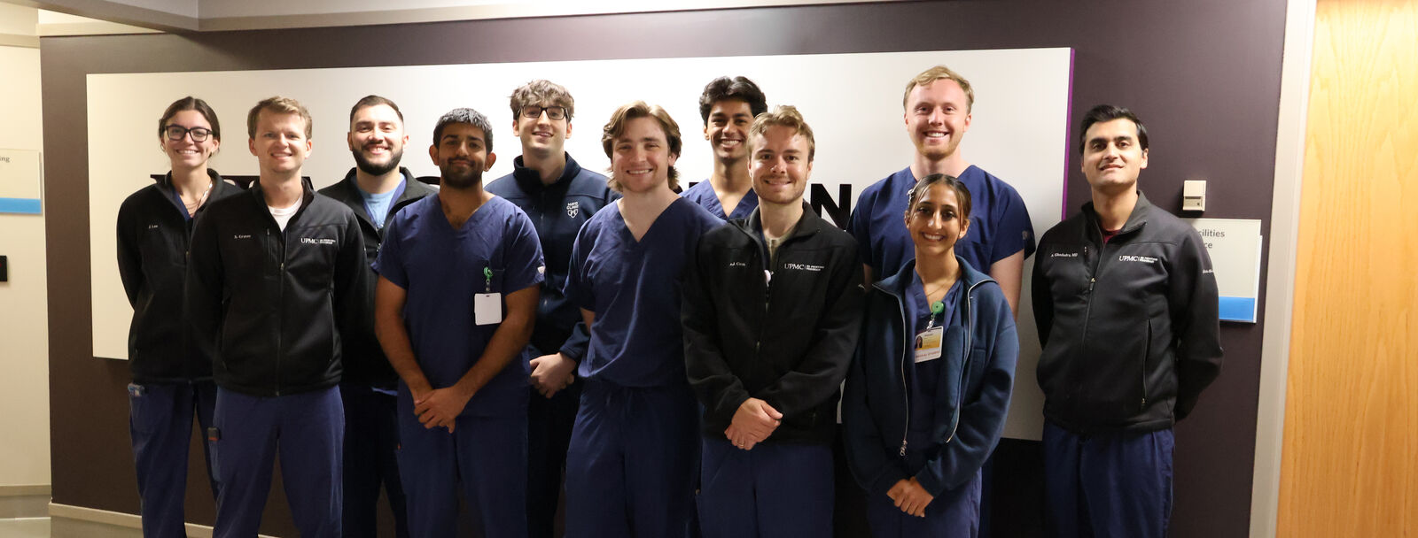
We work on questions that come out of the procedure room.
Interventional radiology treats disease through a small catheter guided by imaging, often in place of open surgery. It is a young enough field that basic questions are still open: which patients do best, how a procedure should be done, and how to tell whether it worked. Our projects start from those questions and run the full length: designing the study, collecting the data, and publishing what we find.
What we are working on
Six kinds of question, at different stages. Some have been running for years; one is just being set up.
-
Studying the outcomes of image-guided therapy
Does the procedure work, for whom, and for how long? Our longest running version of this question is knee embolization, which treats pain by reducing blood flow to inflamed tissue inside the joint. We study who tends to benefit, how the procedure can be made simpler and more comfortable, and how results should be measured so that findings from different centers can be compared.
-
Making sense of rare vascular disease
Conditions too uncommon for any one clinician to see often need described patterns and coordinated care. Hereditary hemorrhagic telangiectasia (HHT) is an inherited condition causing abnormal connections between arteries and veins, which can lead to nosebleeds, anemia, and problems in the lungs, liver, or brain. We work on this and on the wider family of vascular malformations, looking at how they are found on imaging and how care is best organized.
-
Building new procedures from the ground up
Some work starts before there is anything to measure. When kidney function declines without a clear cause, a small tissue sample can point to the diagnosis. We are writing the protocol for random renal biopsy and the safety and quality framework around it now, so the procedure can be studied properly once it is running.
-
Designing devices for one specific patient
Engineering, rather than statistics, is the method here. Growing out of the UPMC 3D Printing Program, this work covers patient-specific anatomic models and surgical guides, newer implant materials, and computer simulation of how an implant is likely to hold up once it is in place.
-
Modeling anatomy at population scale
Large public imaging collections let us study anatomy across thousands of people rather than a handful. We use statistical shape modeling and related methods to describe how anatomy varies from person to person and to look for the patterns that track with disease.
The team
Medical and graduate students work alongside the biomedical engineers of the UPMC 3D Printing Program. The lab meets weekly to go over what moved, what is stuck, and what comes next.
-
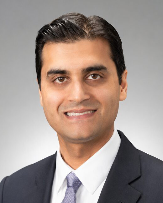 Anish Ghodadra, MD
 -
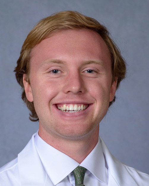 Zachary Spears
 -
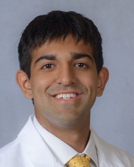 Aizaaz Faiz
 -
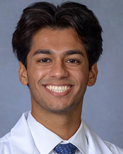 Amogh Vellore
 -
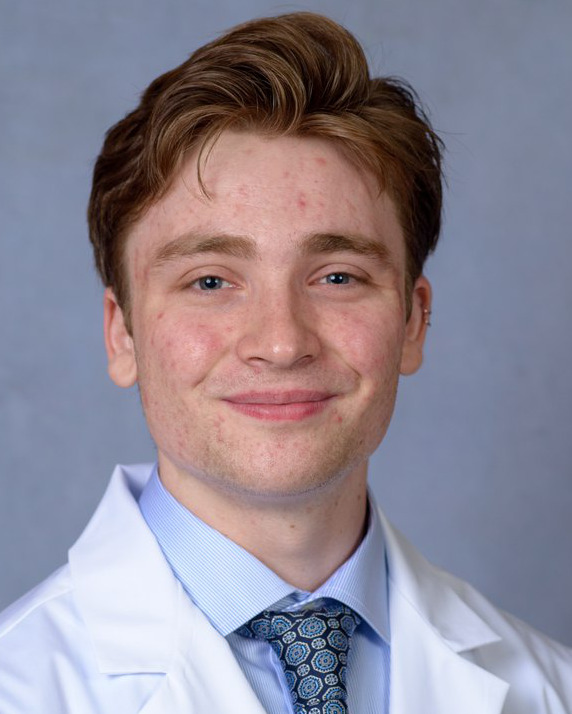 William McKay
 -
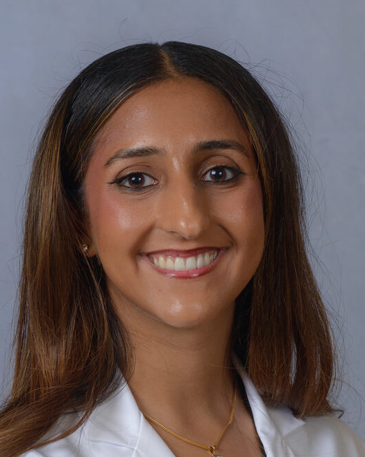 Akarshi Brar
 -
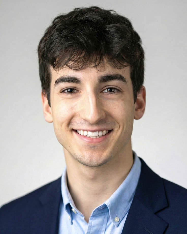 Armani Manov
 -
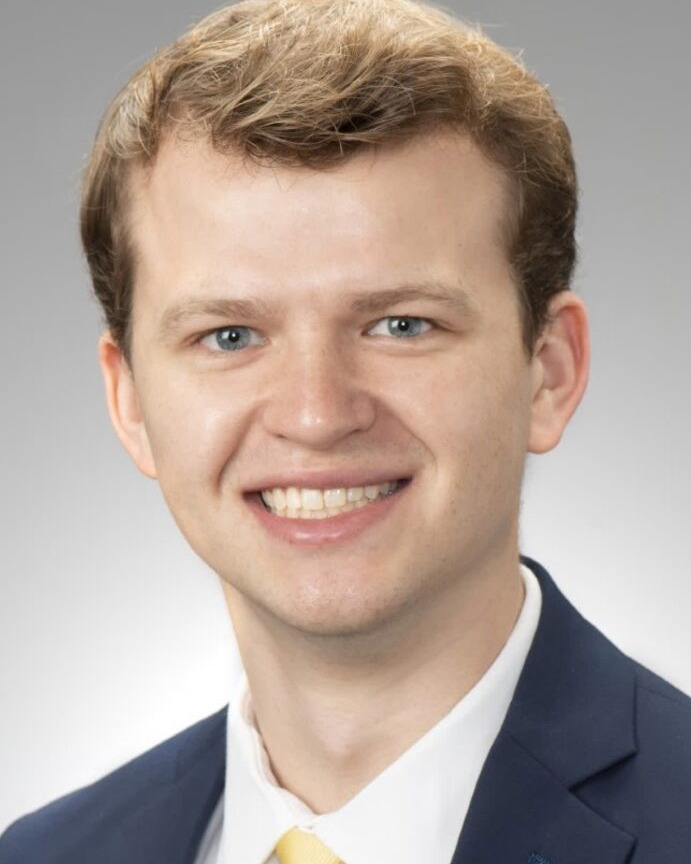 Sean Graves
 -
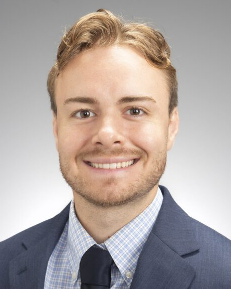 AJ Cook
 -
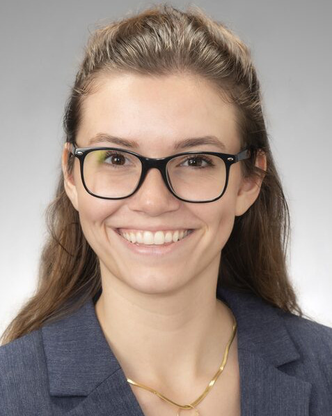 Jadan Law
 -
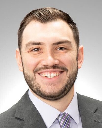 Nicholas Torres
 -
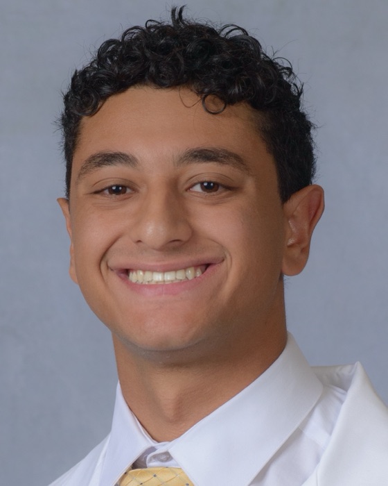 Abrahim Kashkoush
 -
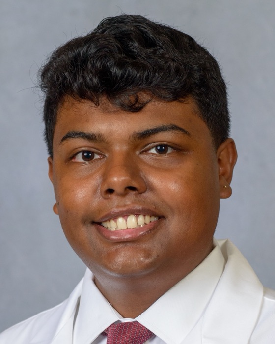 Shreyas Krishnan
Working with us
We take on students who want to see a project through rather than help out at the edges. Collaborations with other groups at Pitt and UPMC, and with centers elsewhere, are welcome. Write to ghodadraa@upmc.edu with a short note about what you are interested in.